Explain how the bones fit together in an articulated skeleton D. Therefore the forearm is in the _____ position.

Coxal Pelvic Bone Posterior View With Labels Appendicular Skeleton Visual Atlas Page 18 Anatomy Flashcards Medical Anatomy Anatomy And Physiology
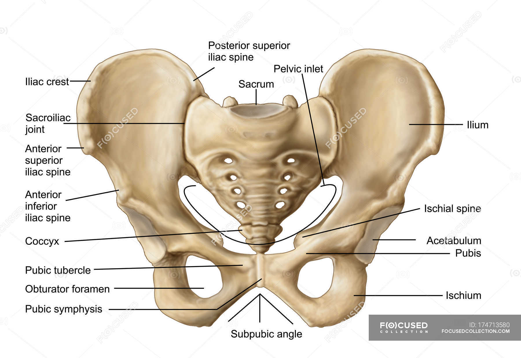
The right and left hip bones also converge anteriorly to attach to each other.
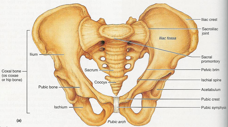
. Label the bone markings on the pelvis. After you have studied the bones in lab label the drawings as a self-test. The pelvic girdle bones and their location relative to each other can be seen in the diagram below.
They will also be described one by one below. Most but not all features you are required to know are shown on the following pages. In the anatomical position the radius and ulna are parallel to one another.
Label the structures on the proximal end of the right femur posterior view. At the front youve got the pubic symphysis. BONES OF THE AXIAL AND APPENDICULAR SKELETON.
There is a printable worksheet available for download here so you can take the quiz with pen and paper. Label the surface features of the right os coxae hip bone medial view. This quiz has tags.
The hip bone has three parts. _____ BIOL 131 - Anatomy Physiology I Lab Exercises 7 8 The Appendicular Skeleton and Joints I. At the back youve got the sacro-iliac joints.
These bones connect the axial skeleton to the lower limbs and therefore play a role in bearing the weight of the upper body. Click on the tags below to find other quizzes on the same subject. The pelvic girdle hip girdle is formed by a single bone the hip bone or coxal bone coxal hip which serves as the attachment point for each lower limb.
Do not spend your. After you have labeled the bones coloring them using the following chart. Each hip bone in turn is firmly joined to the axial skeleton via its attachment to the sacrum of the vertebral column.
Which of the following features is the most proximal feature of the ulna. This is an online quiz called Labeling the Bones of the Skull. Label the bones of the pelvis.
Examine the bones of the pelvic girdle and locate the following. There is a printable worksheet available for download here so you can take the quiz with pen and paper. Figures 176 Pelvic Region and 177 Right Knee STUDY Learn Write Test PLAY Match Created by THagge TEACHER Terms in this set 12 Head of Femur.
Label the surface features of the pelvis. Anatomy and Physiology Part A Drag the appropriate labels to their respective targets. Terms in this set 3 Coxal Bone Hip Bone.
Upgrade to remove ads. The ilium pubis. Correctly label the bones and anatomical features of the fetal skull.
Those are the features of the pelvic bones the hip bones the ossa coxae. Ischium ilium ischial tuberosity greater sciatic notch posterior inferior iliac spine iliac crest posterior superior iliac spine ischial spine lesser sciatic notch obturator foramen acetabulum pubic tubercle ilium symphysis pubis public arch acetabulum sacrum sacral promontory ischium sacroiliac joint. Innominate ilium iliac crest anterior superior iliac spine posterior superior iliac spine greater sciatic notch portion in ischium iliac fossa ischium ischial tuberosity ischial spine lesser sciatic notch pubis pubic symphysis joint between pubic bones pubic arch subpubic angle.
The sacrum articulates above with the fifth lumbar vertebra and below this articulates with the coccyx. Study from the bone list or your textbook after you marked the drawings as instructed on page 6-2. These bones also act as attachments for many muscles and ligaments within the pelvis and lower limbs.
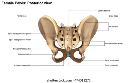
Learn vocabulary terms and more with flashcards games and other study tools. Next Ill just do a quick tutorial on the sacrum and the coccyx. Identify the bones and their landmarks on this posterior view of the pelvic girdle.
The hip bone sacrum and coccyx. Head of Fibula. Label the following bones.
Start studying pelvic girdle label. There are three bones of the pelvis. Identify specific bone markings on the bones of the appendicular skeleton C.
The figure below is an anterior view of the head. Maxilla yellow parietal bone light blue mandible light green temporal bone dark blue nasal bone purple occipital bone dark green frontal bone red zygomatic bone - orange 2. Coxal bone anatomy by bluewoodland 13955 plays 18p Image Quiz.
Label the surface features of the distal end of the femur. Identify the bones of the pelvic and pectoral girdle and their attached limbs. The two pelvic bones are connected anteriorly by the pubic symphysis while posteriorly they articulate with the pelvic spine to form the sacroiliac joints.
THS Anatomy Pelvis Posterior View by feesea 4079 plays 8p Image Quiz. ResetH SacrumMetarsals Tibia FemurPelvic bone Tarsals Fibula. Compare and contrast the.
The pelvis plays several important functions in the human body. About this Quiz. The pelvic spine is the posterior portion of the pelvis below the lumbar spine composed of the sacrum and coccyx.
Ligaments of the Pelvis by RegCaf 3989 plays 11p Image Quiz. This is an online quiz called Label the Pelvis.
Skeleton Pelvis Posterior View 3d Illustration Stock Illustration 474011278
Anatomy Of Human Pelvic Bone With Labels Osteology Biology Stock Photo 174713580
Pelvis Anatomy Recon Orthobullets

Lab 17 Figure 17 1 Pelvis Diagram Quizlet

The Pelvic Girdle And Pelvis Anatomy And Physiology I

Human Skeleton System Pelvis With Labels Anatomy Stock Photo Download Image Now Istock

Pelvic Girdle Posterior View With Labels Appendicular S Flickr

Pelvis Anatomy Anatomy Bones Hip Anatomy
0 comments
Post a Comment